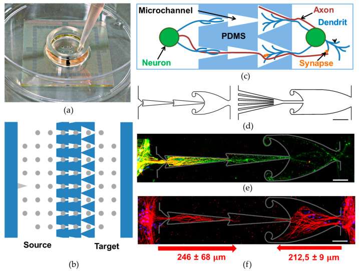
Figure 1.
PDMS chip to grow modular neural networks. (a) Microelectrode array mounted with microfluidic PDMS chip. (b) Schematic view of the microfluidic device mounted to the MEA, with 16 micro-channels connecting the two chambers. Microchannel length was 600 μm in the Fish chip and 700 μm in the Octopus chip. (c) Specific microchannel structure provides unidirectional neurite growth. (d) Schematic view of microchannels in the Fish chip (on the left) and the Octopus chip (on the right), scale bar 100 μm. (e,f) Immunofluorescence images from a hippocampal culture at 21 DIV only in the Source chamber: neurons (b3-tubulin, green), neuronal somas and dendrites (Map2, red) and cell nuclei (DAPI, blue) (e) and in both the Source and the Target chambers—neuronal somas and dendrites (Map2, red) and cell nuclei (DAPI, blue), scale bar 50 μm (f). Chips were removed. Gray lines indicate the manually marked previous locations of the microchannel boundaries. The arrows below indicate the average length of dendrites growing from the Source and the Target modules.

