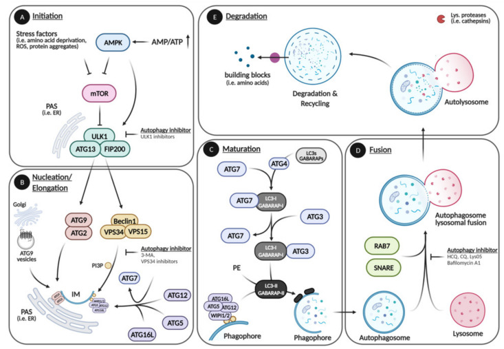
Figure 1.
Autophagic pathway. The autophagic pathway can be divided in five main steps: Initiation, Elongation, Maturation, Fusion, and Degradation. (A) During initiation, stress factors (i.e., low energy, amino acid deprivation, reactive oxygen species (ROS), protein aggregates) activate ULK1 directly or indirectly by inhibiting mTOR. (B) The initiator kinase ULK1 recruits and activates the ATG9–ATG2 complex and the Beclin1 complex that initiate phagophore formation at the PAS. The Beclin1 complex further facilitates membrane elongation by recruiting the first ubiquitin-like conjugation system composed of ATG5, -12, and -16L. (C) During maturation, the second ubiquitin-like conjugation system that includes LC3s (most notably LC3B) and other ATG8 homologs is formed. The ATG8 homologs are cleaved (LC3-I, GABARAP-I) by the ATG4 protease followed by the conjugation with phosphatidylethanolamine (PE) (LC3-II, GABARAP-II) and incorporation into the isolation membrane (IM) via ATG5/12 /16L and ATG3/7 complexes, which leads to the elongation and closure of the autophagosome, thereby incorporating cytoplasmic content. (D,E) When in close proximity, autophagosomes and lysosomes fuse, and the cargo is degraded into cellular building blocks (i.e., amino acids) by lysosomal proteases (cathepsins) via hydrolysis. ULK1 inhibitor: Autophagy-activating kinase 1 inhibitor. 3-MA: 3-Methyladenine. VPS34 inhibitor: PI3K inhibitors. HCQ (Hydroxychloroquine), CQ (Chloroquine), Lys05: Lysosomal lumen alkalizers. Bafilomycin A1 (Baf. A1): Vacuolar type H-ATPase inhibitor. Please see text for further information. Created with Biorender.com, accessed on 3 May 2021.

