Figure 3.
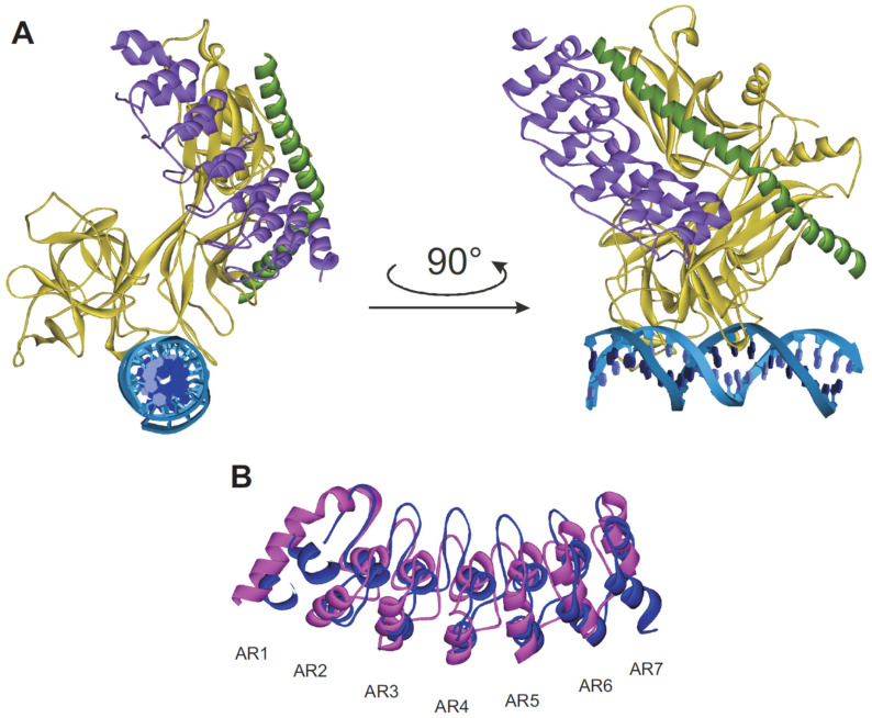
Proposed functional model of Nank. (A) Overall structure of the MAML-1:ANK:CSL:DNA complex (PDB ID: 2F8X) in ribbon representation. The ankyrin domain is purple colored, the MAML-1 polypeptide is colored as dark green and the RHR-N, β-trefoil, and RHR-C domains of CSL are colored gold. The two DNA strands are colored blue and cyan [84]. (B) Superposition of Nank showing the conformational changes induced in the AR1 upon complex formation. Free Nank is depicted in magenta (PDB ID: 1OT8) [82] and Nank in the complexed state in blue (PDB ID: 2F8X) [84].

