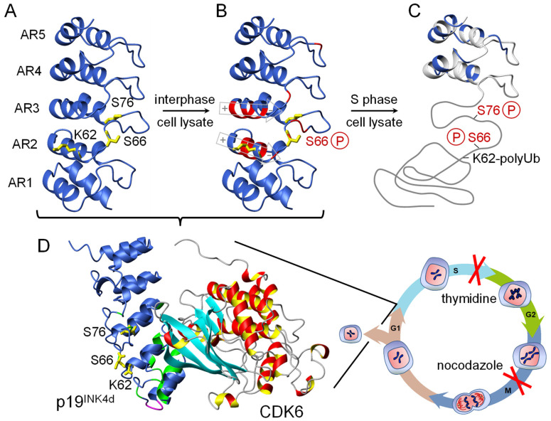
Figure 5.
Cell cycle-dependent conformational changes of ankyrin repeat protein p19INK4d [37]. Phosphorylation at position Ser66 by p38 and at position Ser76 by CDK1 locally unfolds ankyrin repeats 1–3 to release CDK6, which is active during cell cycle progression from the G1 to S phase. Both kinases could only be identified by arresting the cell-cycle after the S phase and the G2 phase. Poly-ubiquitination (polyUb) as the signal for degradation occurs only in the double-phosphorylated state. (A) Residues K62, S66, and S76 are indicated in yellow. (B) Residues with significantly affected backbone NMR chemical shifts upon phosphorylation of S66 are indicated in red. The macroscopic helix dipole moment of helix 4 and 6 is depicted in gray. (C) Only repeats 4 and 5 remain folded after the second phosphorylation of S76, indicated by native backbone chemical shifts (blue). (D) Crystal structure of the CDK6/p19 complex (1BLX.pdb). Green represents residues, which in solution show NMR chemical shift changes upon CDK6 binding.

