Figure 3.

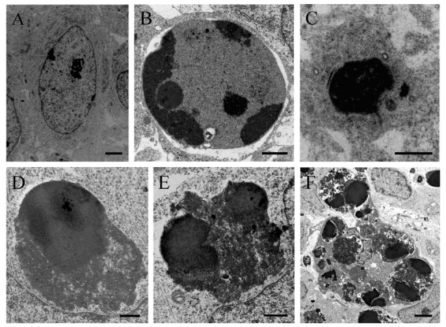
Electron micrograph of a healthy cell (A) and apoptotic cellular profile (B–F) from the neuroepithelium 10 h following BrdU treatment (100 µg/g). (C) Ultrastructural morphology of an apoptotic cell. The masses of compact chromatin display a high electron density and a homogeneous texture. They are associated with an intact nuclear envelope. (D–F) Typical electron-dense apoptotic bodies. (D–E) Examples of apoptotic bodies releasing their contents into the cytoplasm. Copyright © 2021, Wiley. Adapted with permission from Rodríguez-Vázquez, L.; Martí, J. (2000). Administration of 5-bromo-2′deoxyuridine interferes with neuroblast proliferation and promotes apoptotic cell death in the rat cerebellar neuroepithelium. J. Comp. Neurol. 2021, 529, 1081–1096. Scale bar: 2 µm (A), 1 µm (B), 2.5 µm (C), 1 µm (D), 0.5 µm (E), 2 µm (F).
