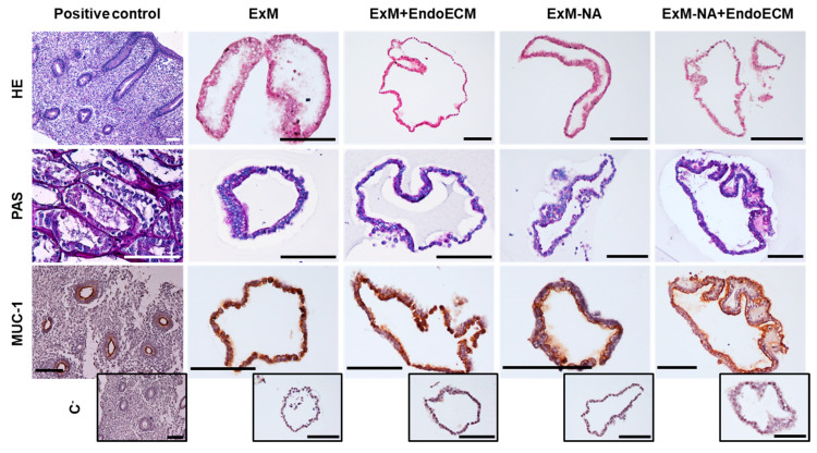
Figure 2.
Characterization of glandular origin in human endometrial organoids by histological and immunohistochemical staining. H&E, PAS staining, and MUC-1 expression in positive controls (endometrium and kidney) and in human endometrial organoids cultured in the four different experimental conditions: ExM, ExM+EndoECM, ExM-NA, ExM-NA+EndoECM. Negative controls for MUC-1 are shown at the bottom of the figure in small size. Scale bars are 100 µm.

