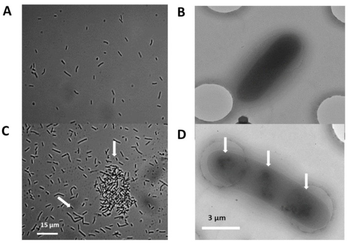
Figure 3.
Micrographs of bacterial cells grown with and without silica nanoparticles (SN): Typical light- and cryo-electron microscopic images of A26 and A26Sfp cells (A,B), and A26SN and A26SfpSN cells (C,D). Arrows indicate cell elongation, bacterial aggregate formation, and changes in nucleoid structure. See Section 4.

