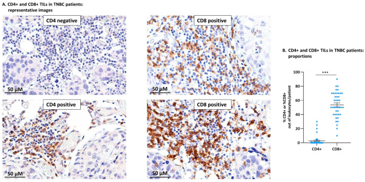
Figure 1.
In TNBC patient tumors, CD8+ TILs dominated the T cell landscape over CD4+ TILs. Biopsy sections of TNBC patients were analyzed for the presence of CD4+ and CD8+ TILs using immunohistochemistry. (A) Images of representative sections stained for CD4 (two different patients: one negative for CD4+ TILs, the second positive for CD4+ TILs) and CD8 (two different patients: both positive for CD8+ TILs). Bar: 50 µm. (B) Semi-quantitative analysis of %CD4+ TILs and %CD8+ TILs out of the leukocyte infiltrate in TNBC patients (n = 41). Data are presented as average ± SE of percentages of the specific subset in all patients. The value of each patient is represented as a dot. *** p < 0.001 (two-tailed unpaired Student’s t-test).

