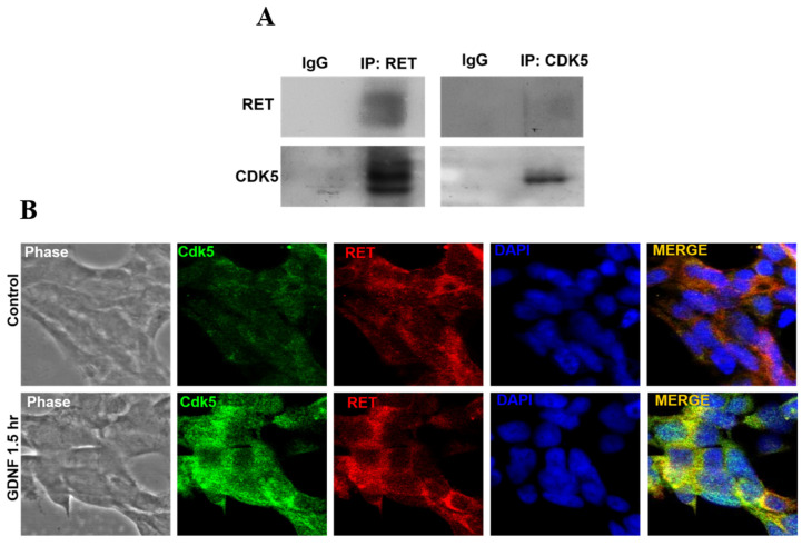
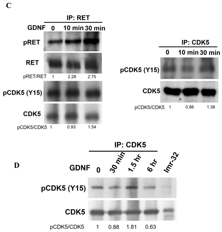
Figure 2.
CDK5 biochemically interacts with RET protein. (A) Western blot data from co-immunoprecipitated RET and CDK5 protein, respectively. RET, CDK5 proteins were detected. (B) Immunocytochemistry data representing intracellular localization of CDK5 (green) and RET (red) proteins with the absence and presence of GDNF. Blue color indicates nucleus staining with DAPI. The yellow color in the merged image indicates the colocalization of two proteins. (C) Western blot data from co-immunoprecipitated RET and protein expression levels of pRET, total RET, pCDK5 and total CDK5 under GDNF treatment for 10 and 30 min, respectively. Western blot data from co-immunoprecipitated CDK5 and the protein expression level of pCDK5 and total CDK5 with GDNF treatment for 30 min, 1.5 h and 6 h, respectively. (D) Western blot data from immunoprecipitated CDK5 and immunoblotting CDK5 and pCDK5 (Y15) under time-dependent GDNF treatment. Imr-32 (human neuroblastoma cell line) serves as a positive control for CDK5 and p35 protein, since their expression are higher in Imr-32 cells.


