Figures 5–8.
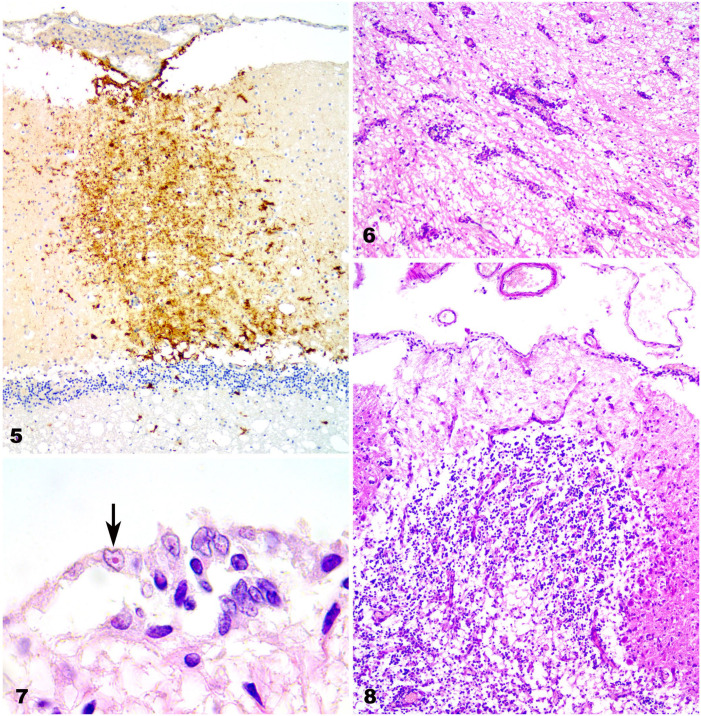
Canine distemper nervous system lesions in vaccinated dogs. Figure 5. CDV-immunopositive cells in the pia matter and throughout the molecular layer in the cerebellar cortex of case 1. IHC against CDV. Original objective 10×. Figure 6. Perivascular lymphocytic cuffing in the cerebrum of case 2. H&E. Original objective 10×. Figure 7. Intranuclear inclusion bodies in ependymal cells (arrow) in the cerebrum of case 4. High magnification of Figure 1. H&E. Original objective 100×. Figure 8. Necrotic area with Purkinje cell loss in the cerebellum of case 2. H&E. Original objective 40×.

