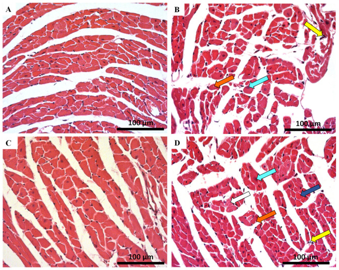
Figure 2.
Cardiac histopathology evaluation done by light microscopy from MTX-treated adult and infant animals and respective controls, as assessed by haematoxylin and eosin staining (experiment 1, mice were euthanized 14 days after the last administration) (A–D). Light micrograph from: (A) infant mice controls in experiment 1, showing normal morphology and structure; (B) infant mice given a cumulative dose of 7.0 mg/kg MTX; (C) adult mice in experiment 1 showed normal morphology and structure; (D) adult mice given a cumulative dose of 7.0 mg/kg MTX. Presence of vacuolization (orange arrow), inflammatory infiltration (yellow arrow), as well as large and uncondensed nucleus (cyan arrow). Cellular oedema (white arrow) and necrotic zones (blue arrow) are evident only in adult mice. Scale bar = 100 µm, n = 3. Images taken at 40× amplification.

