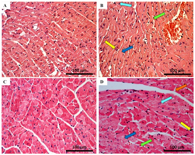
Figure 3.
Cardiac histopathology evaluation done by light microscopy from MTX-treated adult and infant animals and respective, as assessed by haematoxylin and eosin staining controls (experiment 2, adult and infant mice were euthanized 7 or 17 days after the last administration, respectively) (A–D). Light micrograph from: (A) infant mice controls in experiment 2, showing normal morphology and structure; (B) infant mice given a cumulative dose of 6.0 mg/kg MTX; (C) adult mice in experiment 2 showed normal morphology and structure; (D) adult mice given a cumulative dose of 6.0 mg/kg MTX. Presence of vacuolization (orange arrow), inflammatory infiltration (yellow arrow), vascular congestion (green arrow), necrotic zones (blue arrow), as well as large and uncondensed nucleus (cyan arrow) is evident. Scale bar = 100 µm, n = 3. Images taken at 40× amplification.

