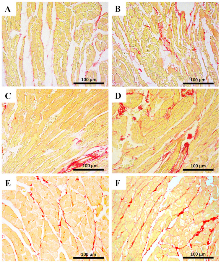
Figure 4.
Sirius red cardiac staining assessed by light microscopy from MTX-treated adult and infant animals and respective controls (A–F). Light micrograph from (A) the control infant mice (experiment 1, mice were euthanized 14 days after the last administration); (B) infant mice injected with a cumulative dose of 7.0 mg/kg MTX; (C) control adult mice; (D) adult mice injected with a cumulative dose of 7.0 mg/kg MTX showing higher fibrosis. (E) Control adults (experiment 2, mice were euthanized 7 days after the last administration); (F) adult mice after receiving a cumulative dose of 6.0 mg/kg MTX. Scale bar = 100 µm, n = 3. Images taken at 40× amplification.

