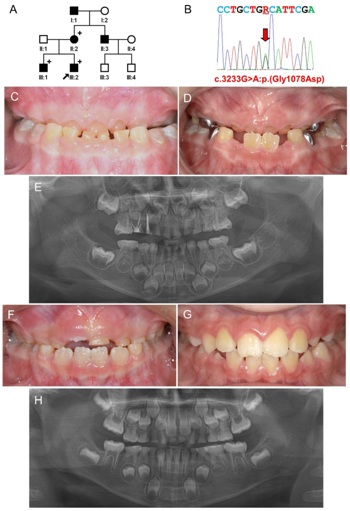
Figure 1.
Pedigree, sequencing chromatogram, clinical photos, and panoramic radiograph of Family 1. (A) Pedigree of Family 1. Symbol filled black indicates affected individual, and the proband is indicated by a black arrow. Plus signs above the symbols indicate participating individuals. Individual ID is shown below the symbols. (B) Sequencing chromatogram of the proband. Nucleotide sequences are shown above the chromatogram. The red arrow indicates the location of the mutation (NM_000089.4:c.3233G>A) (R: A or G). (C) Frontal clinical photo of the proband at age 4 years. (D) Frontal clinical photo of the proband at age 6 years and 7 months. (E) Panoramic radiograph of the proband at age 4 years. (F) Frontal clinical photo of the proband’s brother at age 6 years. (G) Frontal clinical photo of the proband’s brother at age 8 years and 9 months. The incisal edges of the maxillary central incisors were fractured due to an accident. (H) Panoramic radiograph of the proband’s brother at age 6 years.

