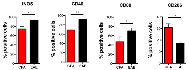
Figure 3.
Immunophenotype of M1 macrophages obtained from CFA and EAE mice. Expression of markers by flow cytometry upon staining cells at cell surface (CD40, CD80 and CD206) or intracellularly (iNOS) in M1 and M2 macrophages obtained from CFA and EAE mice. Data are reported as percentage of positive cells and are representative of eight independent experiments ± SEM * p < 0.05, ** p < 0.01.

