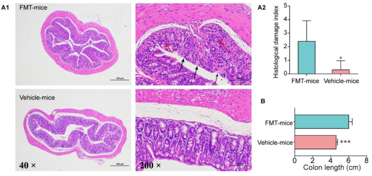
Figure 5.
Inflammatory conditions of the colon after FMT. (A1) Representative images of the colon by H&E staining (40× and 200×). The red arrow indicates morphological changes of mucous layer, and the black arrow indicates edema status in submucosa. (A2) Score of inflammation-associated histological changes in the colon. (B) Colon length after FMT, n = 5. Data are means ± SD and analyzed by Student’s t-test. * p < 0.05, *** p < 0.0001.

