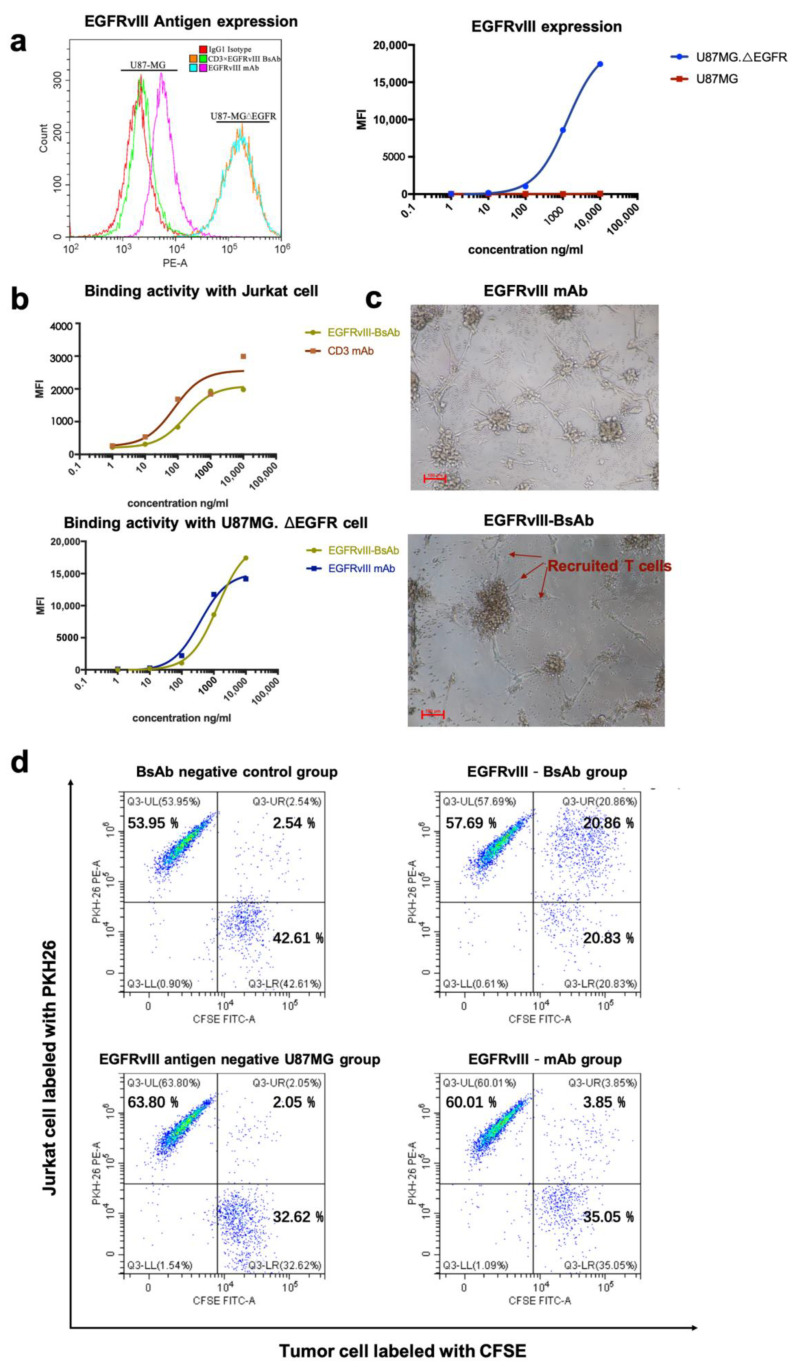
Figure 3.
(a) EGFRvIII antigen expression level on mutant GBM cell line U87MG.ΔEGFR and wild-type GBM cell line U87MG. (b) A binding activity comparison of EGFRvIII-BsAb and CD3 mAb with Jurkat cells (CD3-positive) (upper), as well as a binding activity comparison of the EGFRvIII-BsAb and the EGFRvIII mAb with U87MG.ΔEGFR cells (EGFRvIII-positive) (lower). (c) Photographs of the redirection of T cells to cancer cells by 0.01 ng/mL EGFRvIII-BsAb or EGFRvIII mAb. (d) FACS analysis of the redirection of CD3+ Jurkat cells to cancer cells by EGFRvIII-BsAb. Jurkat (CD3+) cells labeled by PKH26 (PE-A), as well as U87MG.ΔEGFR cells labeled by CFSE (FITC-A).

