Figure 1.
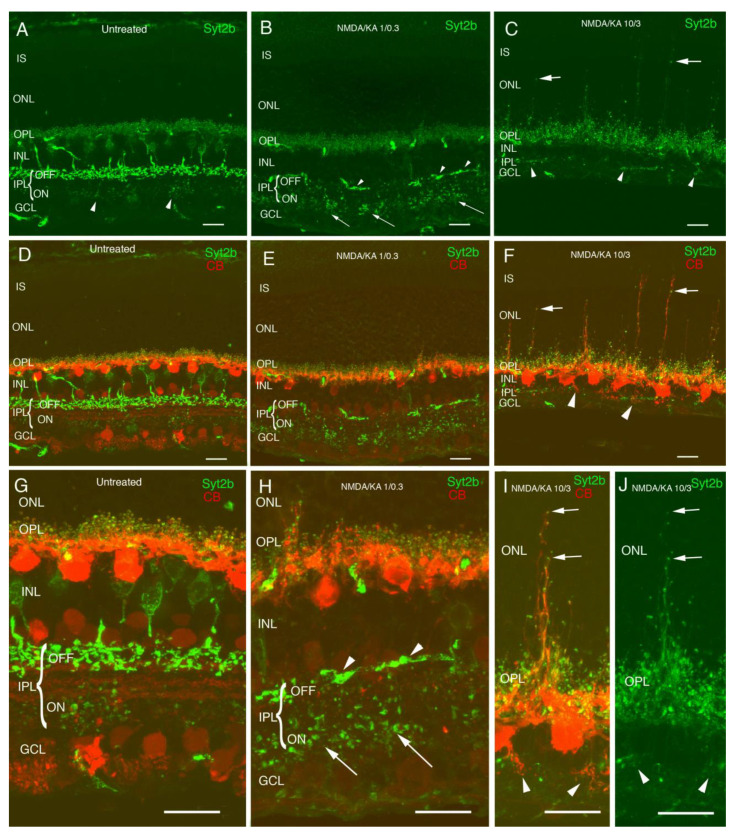
Morphological changes in OFF cone bipolar cells and horizontal cells in N-methyl-DL-aspartate (NMDA)/kainic acid (KA) treated retinas. Cryostat cross-sections immunostained with antibodies against Syt2b showing OFF and ON cone bipolar cells (A). Axon terminals of type 2 cone bipolar cells can be observed in the stratum of the OFF and those of type 6 cone bipolar cells stratify in the stratum of the ON showing less immunoreactivity intensity (A,D arrowheads). Double immunolabeling with calbindin antibodies shows dendrites and axon terminals of horizontal cells in the OPL (D,G). NMDA/KA 1/0.3 treated retinas show OFF cone bipolar cell death, and only a few distorted axons remain in the stratum of OFF (B,E,H arrowhead). In contrast, axon terminals of the ON cone bipolar cells can still be identified in the inner plexiform layer (IPL) stratum ON (B,H arrows). At this dose, the terminals of the horizontal cells show a normal morphology, and only some sprouting can be observed (E,H). NMDA/KA 10/3 treated retinas induce a clear reduction of the inner retina. While the INL, IPL, and GCL layers show a drastic thickness decrease, the IS, ONL, and OPL layer thickness are maintained, indicating that photoreceptors are not affected by the treatment (C,F). Neither ON nor OFF bipolar cells can be detected (C,F) in this condition, and horizontal cells display abnormal dendrite sprouting towards the ONL (C,F,I,J arrows) and the IPL (C,F,I,J arrowheads). IS—inner segments; ONL—outer nuclear layer; OP—outer plexiform layer; INL—inner nuclear layer; IPL—inner plexiform layer; GCL—ganglion cell layer. All of the images were obtained from temporal retina, ca. 500 µm from the optic disk. Scale bar of 10 µm.

