Figure 3.
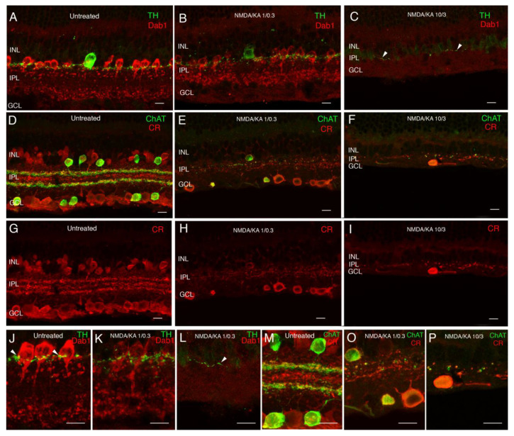
Response of amacrine cells to excitotoxicity. Double immunolabeling with antibodies against tyrosine hydroxylase (TH) and Dab1 (A–C,J–L). Tyrosine hydroxylase shows the dopaminergic amacrine cells and their dendritic plexus in the S1 stratum in the IPL (A,J, green). Dab1 shows the AII amacrine cells, whose typical lobular appendages are mainly in the OFF layer and their dendritic terminals in the ON layer (A,J red). Synaptic contacts from dopaminergic cells around the cell bodies of AII amacrine cells can be observed (J arrowheads). A decrease in TH and Dab1 immunoreactivity intensity is found in response to the 1/0.3 concentration of NMDA/KA. The morphology of AII amacrine cells looks disorganized, but the synaptic contacts with the dopaminergic cells still remain (B,K). At the NMDA/KA 10/3 dose, AII amacrine cells cannot be identified and only a few dendrites of dopaminergic cells can be observed in the S1 stratum of the IPL (C,L arrowhead). Double immunolabeling with antibodies against calretinin and choline acetyltransferase (D–I,M–P). Calretinin immunoreactivity labels several types of amacrine cells and ganglion cells with three typical plexuses of dendrite stratification in the IPL (D,G,M red). ChAT immunoreactivity is found in starburst amacrine cells, whose cell bodies are located in the INL and in the ganglion cell layer, and their dendrites stratify in two specular plexuses in the ON and OFF layers of the IPL (D,M green). At the 1/0.3 concentration of NMDA/KA, calretinin immunoreactive amacrine cells cannot be identified, and only a few ChAT amacrine cells and some CR ganglion cells remain (E,H,O). Both plexuses experience a big disruption and disorganization. At a 10/3 concentration, only some spots of ChAT and CR immunoreactivity can be observed in the IPL, accompanying IPL degeneration (F,I,P). INL—inner nuclear layer; IPL—inner plexiform layer; GCL—ganglion cell layer. Scale bar of 10 µm.

