Figure 7.
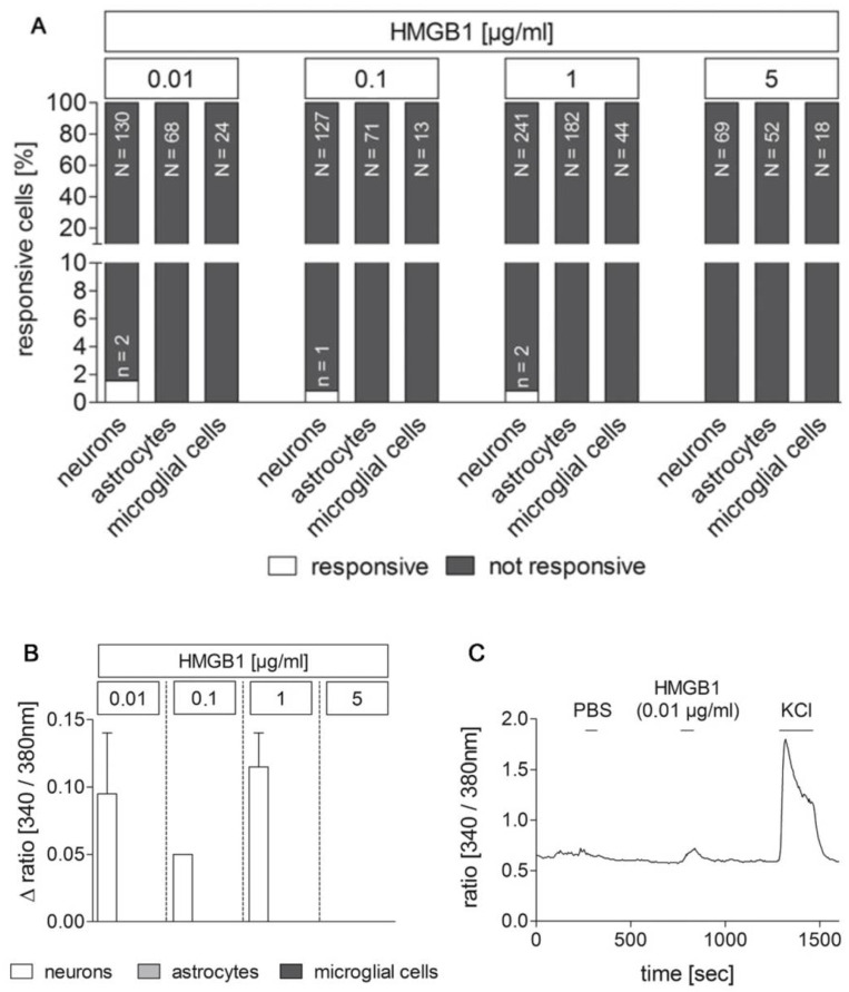
Responses of cultured area postrema (AP) cells upon stimulation with HMGB1 in Ca2+ imaging experiments. Primary cell cultures of the rat AP were used for Ca2+ imaging experiments 4–5 days after preparation. HMGB1 was applied in serial dilutions (0.01, 0.1, 1, 5 µg/mL) to identify stimulus-induced changes in the ratio [340/380 nm] indicative for changes in intracellular calcium concentrations [Ca2+]i. Cell types were characterized by immunocytochemistry after each experiment. A rather small population of neurons responded to stimulation with low doses of HMGB1 with an increase in [Ca2+]i, while astrocytes and microglial cells did not show any response ((A); n = responsive cells, N = total amount of investigated cells). Mean Δratios [340/380 nm] ± SEM of responsive cells are shown in (B). A representative example of an HMGB1 responsive cell is presented in C. PBS was applied as control, while KCl (50 mM) was used to confirm neuronal viability. The numbers of investigated cultures and independent experiments are provided in Table 2.

