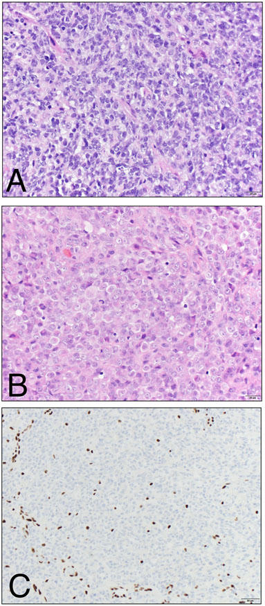
Figure 2. Histological features of ATRT.
A) The hypercellular tumor is usually predominantly composed of primitive-appearing cells with scant cytoplasm and hyperchromatic nuclei. Mitotic figures, apoptotic bodies and necrosis may all be readily identified. B) A subset of cells may show abundant, globular eosinophilic cytoplasm, reminiscent of rhabdoid cells. C) SMARCB1 (INI1 / BAF47 / SNF5) immunohistochemistry shows uniform loss of expression in the tumor cells, while expression is retained in endothelial nuclei serving as positive internal controls. (A, B – Hematoxylin and Eosin stain, 400x magnification; C – BAF47 clone (BD Biosciences), 200x magnification).

