Figure 6.
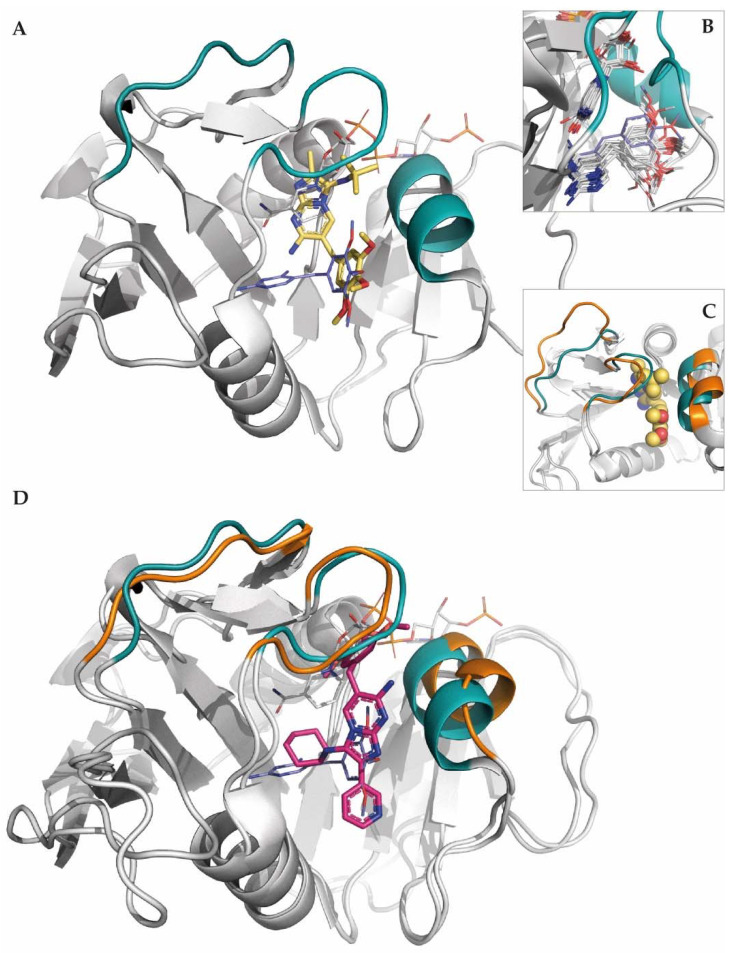
Binding mode of 1a to the DHFR of E. coli. (A) The docked positions of 1a and TMP are shown with their C atoms depicted as yellow sticks and in deep blue, respectively, and with the C atoms of NADPH in gray. (B) Superposition of the crystallographic poses of TMP (C atoms in gray). The crystallographic structure of TMP in PDB entry 2W9H, in which the trimethoxybenzene group sterically collides with the nicotinamide ring of NADPH, is shown in deep blue. (C) Loops Met20 and F-G, and helix 3 are highlighted to illustrate the differences in the 3D structures of PDB entries 3DAU (turquoise) and 4KM2 (orange). (D) The docked position of 1b (C atoms in magenta) in a homology model of E. coli DHFR built using the open structure 4KM2 as a template. The position of TMP and NADPH is shown with their C atoms depicted as deep blue and gray, respectively.

