
Figure 1.
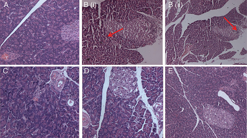
Histopathological changes in the pancreas. A) Normal control group rats with no damage to acinar cells; B (i) and B (ii) disease control group: L-arginine treated rats with vacuolar degeneration and extensive damage of the acinar cells with infiltration of leucocytes-red arrow; C) positive-control melatonin group of rats with normal architecture of acinar cells; D, E) GSAE-treated rats showing mild damage to the normal echotexture of acinar cells
GSAE: Granny Smith apple extract