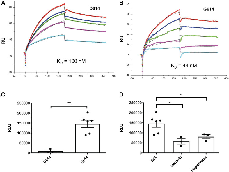
FIGURE 5.
The SV2-S variant G614 protein has a higher binding affinity to heparin than the wildtype SV2-S D614 protein, and the entry of SAS-Cov-2 G614 pseudotyped virus into host cells depends on cell surface HS and can be inhibited by heparin. (A,B) SPR binding kinetics sensorgrams between heparin and SV2-S D614 or SV2-S G614. Concentrations of D614 were (from top to bottom): 1,000, 800, 600, 400, and 200 nM, respectively (A), and G614 were (from top to bottom): 1,000, 600, 400, 200, and 100 nM, respectively (B). The black curves are the fitting curves using a 1:1 Langmuir model from BIAevaluate 4.0.1. (C) Infection of 293T-ACE2 cells by the same titer of SARS-Cov2-D614 and SARS-Cov2-G614 pseudovirus. (D) SARS-Cov2-G614 pseudovirus depends on HS to infect host cells. The 293T-ACE2 cells were infected with SARS-Cov2-G614 pseudovirus expressing luciferase after the cells were treated with heparinases I-III (5 mU/ml) or in the presence of heparin (2 μM). The luciferase activity was measured 48 h after the virus infection. VSV pseudotyped virus was used as a control to make sure each well has comparable cell numbers (data are not shown). The experiments were repeated at least 3 times and an unpaired t-test was performed for two-group comparison. Data are presented as mean ± SD. *p < 0.05, **p < 0.01.

