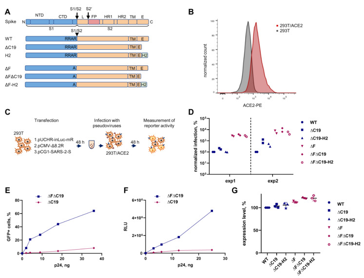
Figure 1.
Development of a SARS-CoV-2 cell-free infection test with PVs. (A) A schematic illustration for S-protein variants used in pseudovirus infection tests. Six different constructs of the S protein were generated by PCR mutagenesis. (B) Evaluation of the ACE2 surface expression on 293T cells stably transduced with the hACE2 using flow cytometry. (C) Experimental setup for SARS-CoV-2 cell-free infection measurement. (D) The levels of infection detected with different variants of the S protein. PVs were added to 293T/ACE2 cells in an equal amount based on HIV-1 Gag quantification. The luciferase activity measured for a mutant spike was normalized to that obtained for the wild-type S protein. Two independent experiments with two different PV preparations were performed. (E,F) The levels of cell-free infection with indicated PVs were measured using either GFP (E) or inLuc (F) reporter. (G) The levels of S protein expression on PV-producing cells estimated by flow cytometry. To express indicated variants of protein S, 293T cells were transfected and stained with convalescent human serum in 48 h. Median fluorescence intensity (MFI) level was calculated for every mutant in the gate of transfected cells and normalized to the MFI detected for wild-type S protein.

