Figure 7.
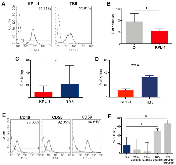
In vitro effect of anti-PSGL-1 antibodies on ALCL cell lines. (A) SU-DHL-1, a representative ALCL cell line, was analyzed by flow cytometry using two independent anti-PSGL-1 antibodies, KPL-1 and TB5, and a FITC-conjugated secondary antibody. (B) KPL-1 antibody was able to partially reduce SU-DHL-1 adhesion on EA.hy926 endothelial cell lines. (C) TB5 is able to induce direct cytotoxicity, analyzing residual cell viability after 48 h of incubation with SU-DHL-1 cells. (D) TB5, more than KPL-1, is able to induce ADCC of SU-DHL through PBMC activation and analyzing LDH release. (E) SU-DHL-1 cells evidenced the expression of CD46, CD55 and CD59 in FACS analysis after the incubation with specific antibodies and FITC-labeled secondary antibodies. (F) The binding of TB5 on the surface of ALCL cells is effective in inducing low levels of complement-dependent cytotoxicity using human serum as the source of complement. The percentage of lysed cells through CDC rises to significant values following blockage of the membrane complement regulatory proteins CD46, CD55 and CD59 expressed on SU-DHL-1 cells. p-values: * ≤ 0.05; *** ≤ 0.001.

