Figure 3.
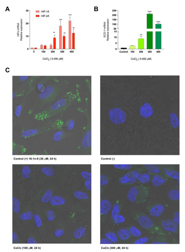
Chemical hypoxia promotes SCD-1 expression in vitro. (A) Caki-2 cells were exposed to different concentrations of CoCl2 (0–400 µM) for 24 h, and the development of the hypoxic microenvironment was tested with the expression of HIF-1A and HIF-2A by RT-qPCR. (B) Then, SCD-1 overexpression was detected under the same experimental conditions by RT-qPCR. (C) The evaluation of LD formation was determined by confocal microscopy. Caki-2 cells were exposed to 18:1n-9 (30 µM) for 24 h as a positive control for LD formation. Magnification 400×. Data are expressed as the means ± SEM and are representative of three independent experiments. ** p < 0.01, *** p < 0.001 and **** p < 0.0001, significantly different from the control.

