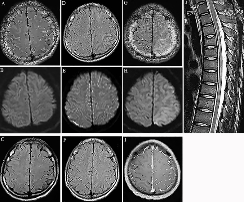
Figure 1.
(A) Fluid attenuation inversion recovery (FLAIR) image showed hyperintensity in the cortical region of right frontal and parietal lobes without white matter involvement (Case 1). (B) Diffusion weighted imaging (DWI) showed no obvious signal change in the cortex (Case 1). (C) MRI scan showed the abnormality disappeared at the follow-up scan (Case 1). (D) Hyperintensity in the cortical regions of left parietal lobes in MRI FLAIR images 7 years before this episode (Case 1). (E) DWI showed no obvious signal change in the cortex (Case 1). (F) MRI scan showed the abnormal lesions disappeared at the follow-up scan (Case 1). (G) Hyperintensity in the cortical regions of left frontal and parietal lobes in MRI FLAIR images (Case 2). (H) DWI showed mild hyperintensity in the involved cortical regions (Case 2). (I) Gadolinium enhanced T1-weighted image showed meningeal linear enhancements in the sulci of left brain lobes (Case 2). (J) T2-weighted image showed hyperintensity in the spinal cord from thorax 2 to 9 segments. (Case 2).

