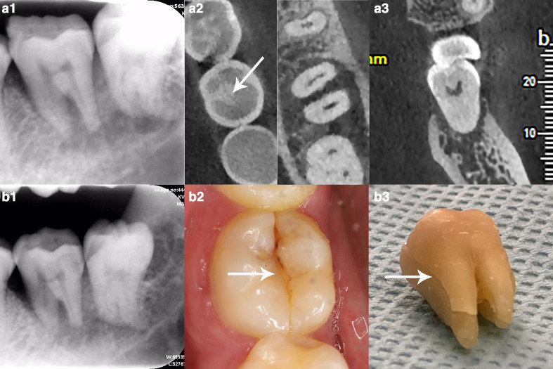
Figure 2.
A case of cracked tooth progressing to split tooth (tooth 36). (a1) Periapical radiograph (PR) showing extensive bone resorption around the root. (a2) Axial image showing a mesiodistal hair-like hypodense line (arrow) that is present on the crown but disappears on the root. (a3) Coronal reconstruction image showing severe buccal and lingual alveolar bone resorption around the root. Cracked tooth with acute inflammation was diagnosed after an incision was made on the periodontal abscess; however, the patient did not undergo further treatment. (b1) PR taken 1 year after the primary referral at our hospital showing more extensive bone resorption around the root. (b2) An obvious mesiodistal fracture line (arrow) can be seen on the occlusal surface. (b3) The extracted tooth has split completely (arrow).

