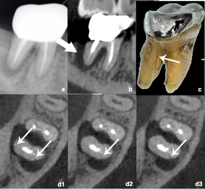
Figure 5.
(a) PR showing a low-density shadow around the distal root of tooth 46. (b) Sagittal reconstruction image showing alveolar bone resorption around distal root (arrow). (c) The extracted tooth showing the fracture line (arrow). (d1–d3) Subtle irregular hypodense line (arrows) on the distal root without displacement of the two fracture segments.

