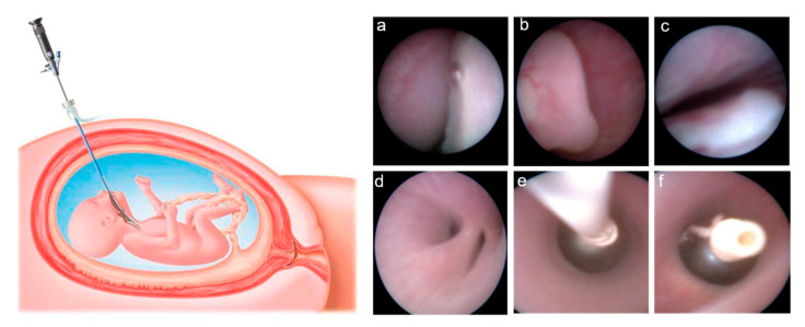
Figure 1.
On the left side of the figure, schematic drawing showing intrauterine access to the fetal trachea by maternal percutaneous insertion of a fetoscope (reproduced from: Deprest, J.; Gratacos, E.; Nicolaides, K.H. Fetoscopic tracheal occlusion (FETO) for severe congenital diaphragmatic hernia: Evolution of a technique and preliminary results. Ultrasound Obstet. Gynecol. 2004, 24, 121–126 [11]). On the right side, endoscopic images showing the fetal oral cavity (a), the epiglottis (b), and the vocal cords (c), through which the fetoscope is advanced to reach the carina above the main bronchial bifurcation (d). At this level, the balloon is released through a catheter (e), inflated and detached to obtain tracheal occlusion (f).

