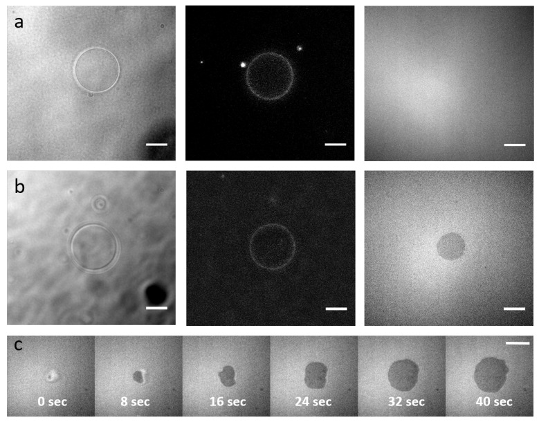
Figure 6.
Functionality of the integrins. (a,b) pGUVs containing integrins are deposited on Supported Lipid Bilayers (SLBs) without RGD ligand of integrins (a) or with RGD (b). Left: Bright-field channel, GUVs. Middle: Epi-fluorescence channel showing integrin incorporation in the GUV. Right: RICM, dark regions indicate adhesion. (c) Timelapse of an integrin containing pGUV adhering to an RGD functionalized SLB (time is indicated). Scale bars: 10 m.

