Figure 5.
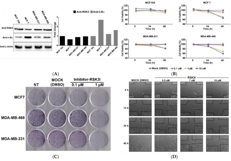
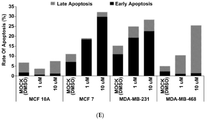
RSK3I inhibits tumorigenesis and increases apoptosis in breast cancer cells. (A) The basal expression levels of RSK3 and IκBα in breast cancer and normal breast epithelial cell lines were analyzed using Western blots. Representative results from two experiments are shown. (B) Cell viability was analyzed using a CCK assay. RSK3I was added at concentrations of 0.1 μM, 1 μM, and 10 μM for 24 h. (C) Foci assays were performed to analyze the growth of breast cancer cells (MCF7, MDA-MB-468, and MDA-MB231). Cells were incubated with RSK3I for 14 days and stained with 0.5% crystal violet. Cell counting was performed using the ImageJ (Java-based image processing program) tool. (D) Wound-healing assays were performed to analyze the effect of RSK3I on the migration of MDA-MB-231 cells. The cells were treated with several concentrations (0.1 μM, 1 μM, and 10 μM) of RSK3I, and cell migration was monitored for 48 h. (E) The effect of RSKI on apoptosis was assessed by Annexin V/PI staining followed by flow cytometry analysis. RSK3I was added to breast cancer cells (MCF7, MDA-MB-468, and MDA-MB-231) or a normal breast epithelial cell (MCF10A) at concentrations of 1 μM and 10 μM for 24 h, and DMSO was used as a control.


