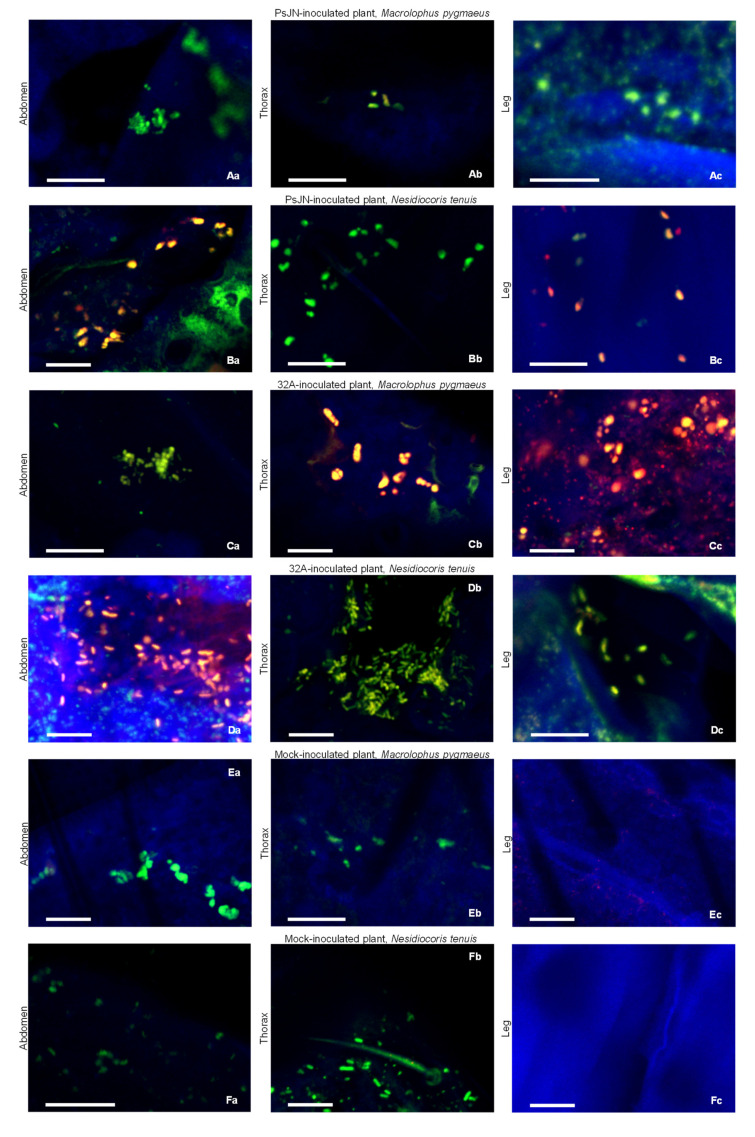
Figure 2.
Location of endophytic bacterial strains on mirids. Macrolophus pygmaeus (A,C,E) and Nesidiocoris tenuis (B,D,F) abdomen (a), thorax (b) and leg (c) samples were collected at the end of the acquisition period (Day 11) on plants inoculated with Paraburkholderia phytofirmans PsJN (PsJN) (A,B) or Enterobacter sp. 32A (32A) (C,D) or mock-inoculated plants (E,F). PsJN cells were hybridised with the EUBmix and Bphyt probes (A,B) and 32A cells were hybridised with the EUBmix and Gam42a probes (C,D). Mirids fed on mock-inoculated plants (E,F) were hybridised with the EUBmix and Gam42a probes. Five replicates (mirids) were analysed for each treatment and representative pictures were selected. Bars correspond to 10 µm.

