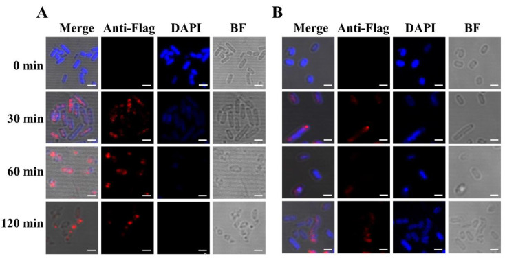
Figure 3.
Immunofluorescence staining of lysis protein EM-Flag. BL21(DE3) bacterial cells containing pLysS and pET28a-EM-Flag (A) and BL21(DE3) bacterial cells only containing pET28a-EM-Flag (B) were induced by IPTG, and cells were collected at 0 min, 30 min, 60 min, and 120 min. In these bacteria cells, nucleic acid was non-specifically bound and labeled by the DPAI blue stain. The Flag tag at the C-terminus of lysis protein EM was specifically recognized and bound by the mouse anti-Flag tag of the primary antibody. The Alexa Fluor® 647 (red)-conjugated goat anti-mouse secondary antibody was specifically bound to the primary antibody. Bar: 2.0 µm.

