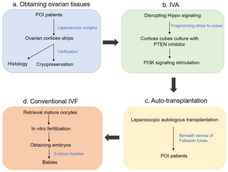
Figure 4.
Schematic diagram of IVA. (a) Obtaining ovarian tissues. Under laparoscopic surgery, ovaries were obtained and cut into strips for histology and cryopreservation after vitrification. (b) IVA. After thawing of cryopreserved ovarian strips, the strips were fragmented into 1–2 mm3 cubes and cultured with Akt stimulators for 2 days. (c) Auto-transplantation. After two days of culture, the ovarian cubes were autografted beneath the serosa of the fallopian tubes. (d) Conventional IVF. Follicle growth was stimulated by injection of FSH. When preovulatory follicles were found, mature oocytes were retrieved for IVF to obtain embryos.

