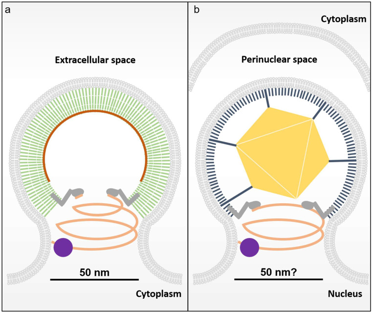
Figure 2.
The process of HSV-1 nuclear egress and scission resembles the retroviral egress pathway. (a) Diagram of the well characterized HIV-1 budding site. HIV-1 Gag (green sticks) binds RNA (dark orange) and assembles on the membrane and recruits ALIX (dark grey) via the L-Domain located in its C-terminal p6 domain. HIV-1 budding necks have been shown typically to adopt a diameter of approximately 50 nm prior to membrane scission and this topology is likely to be shared by the NEC given the mechanistic constraints of the ESCRT-III membrane scission machinery. The downstream ESCRT-III is shown in salmon and the VPS4 membrane scission machinery in purple. (b) HSV-1 particle assembly begins in the nucleus where a newly formed capsid must transit the nuclear envelope by assembly and budding. Schematic of the HSV-1 nuclear egress complex prior to membrane scission assembled with its capsid as it buds through the nuclear envelope. A single virus capsid is shown as a gold hexagon and the NEC UL31-UL34 heterodimer is shown as membrane-associated sticks (black) that occasionally interact with the vertices of the capsid. ALIX is recruited to sites of nuclear egress through interactions with NEC and mediates the recruitment of the downstream ESCRT components to facilitate membrane scission.

