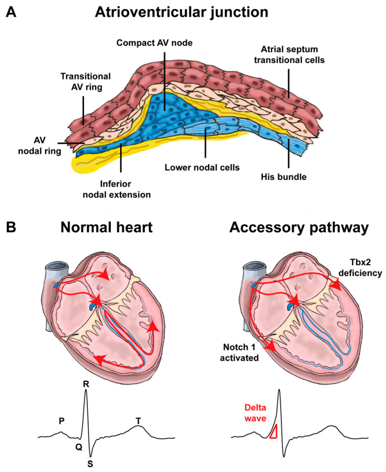
Figure 4.
Atrioventricular junction and impulse propagation (A). Schematic representation of the different components of the atrioventricular junction (Inspired by [53,70]). Note that lower nodal cells share a common origin with the His bundle cells. (AV: atrioventricular) (B). In normal hearts, the electric impulses initiated by pacemaker cells in the sinoatrial node propagate through the atrial myocardium and trigger its contraction. At the atrioventricular node, the impulses are delayed for a period to facilitate alternating contraction of the atrial and ventricular myocardium. After the atrioventricular delay, the electrical impulses rapidly travel to the ventricular myocardium via the His-Purkinje system and stimulate the ventricular myocardium. In Notch1-activated and Tbx2-deficient hearts, accessory pathways are formed as a result of malformation of the atrioventricular canal myocardium, commonly right-sided in Notch1-activated mice and left-sided in Tbx2-deficient mice. Because of faster conduction through the accessory pathways than through the atrioventricular node, the ventricular myocardium is prematurely stimulated (preexcitation). The ECG shows a short PR interval, a slurred upstroke (“delta wave”) of the QRS complex and a widened QRS complex.

