Figure 2.
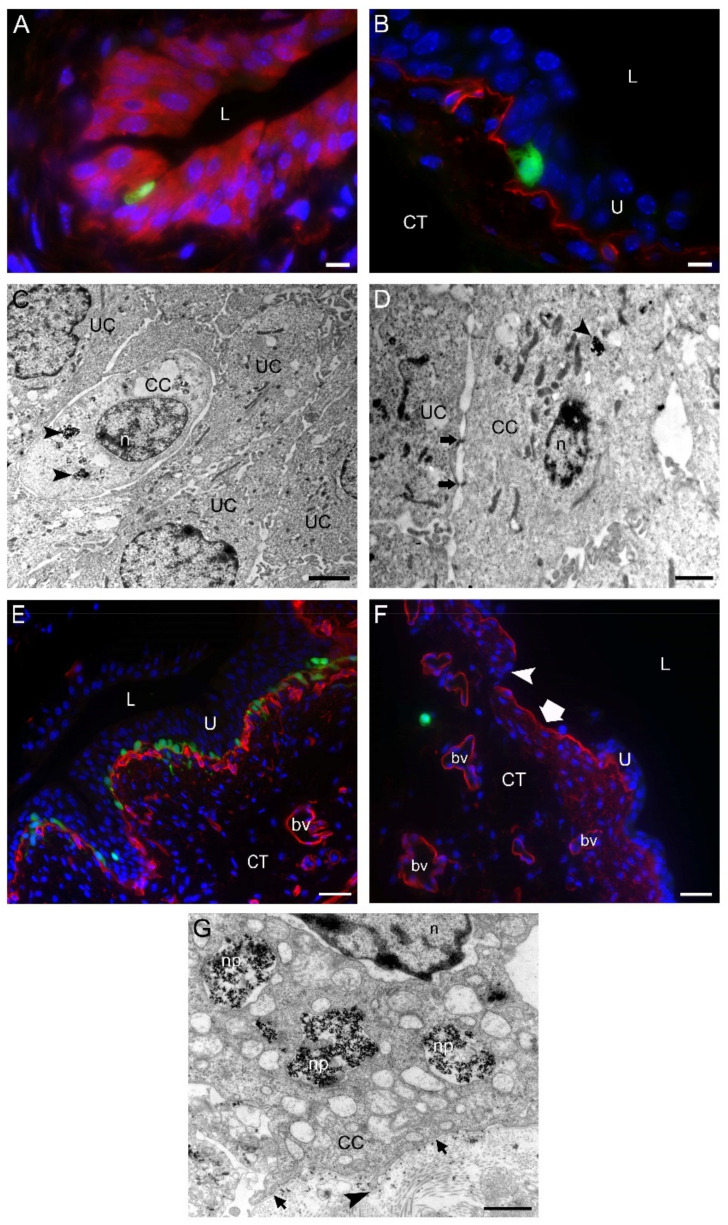
One to three days after intravesical application of MB49-GFP cancer cells (CC) with internalized nanoparticles. (A) An MB49-GFP CC (green fluorescence) migrating through the urothelium toward the basal lamina. Urothelial cells were immunolabeled for keratin 7 (red fluorescence). The dark spots in a CC are endosomes with metal nanoparticles. (B) An MB49-GFP CC (green fluorescence) reaching the basal lamina immunolabeled for collagen IV (red fluorescence). (C,D) Transmission electron micrographs of migrating CC with endocytosed nanoparticles (arrowheads). CC during their trans-urothelial migration form desmosomes (arrows in (D)) with urothelial cells (UC). (E) The region of the urinary bladder wall where numerous MB49-GFP CC (green fluorescence) after the migration through the urothelium are reaching underlying basal lamina immunolabeled for collagen IV (red fluorescence). (F) Individual MB49-GFP CC (green fluorescence) penetrating the basal lamina (red fluorescence due to immunofluorescence labeling of collagen IV) and appearing in the connective tissue. Note the area of the urothelium that is completely peeled off (thick arrow), and the part of degraded basal lamina (arrowhead). (G) A higher magnification view of the part of the CC with internalized nanoparticles (np) close to the basal lamina (arrows). Arrowhead denotes the invadopodium protruding through the degraded part of the basal lamina. Nuclei were stained with DAPI (blue fluorescence) in (A,B,E,F). U—urothelium; L—lumen of the urinary bladder; CT—connective tissue; n—nucleus; bv—blood vessel. Bars are 10 µm (A,B); 2 µm (C), 1 µm (D,G); and 50 µm (E,F).

