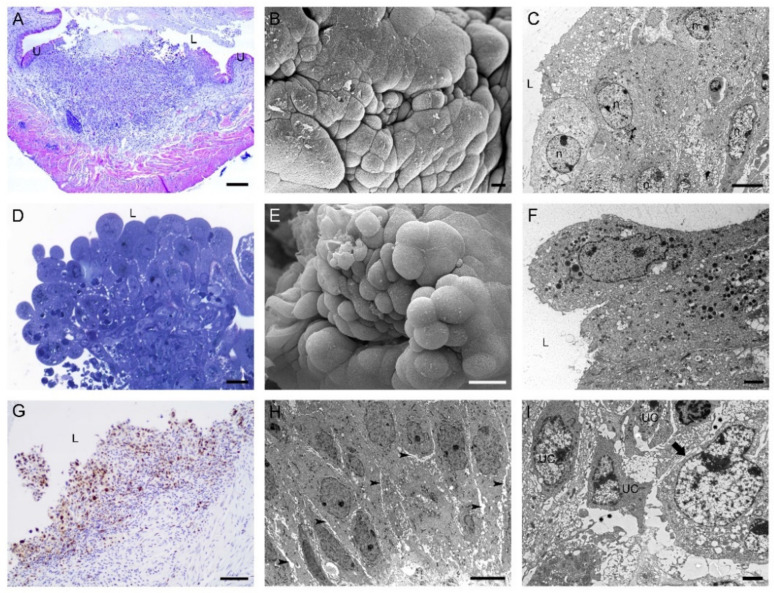
Figure 5.
Characteristics of tumors developed in mouse urinary bladders 3 weeks after intravesical application of MB49-GFP cancer cells. Histological and ultrastructural features of sessile bladder tumors in a (A) histological section (H&E staining), (B) scanning electron micrograph, and (C) transmission electron micrograph. Histological and ultrastructural features of papillary tumors in a (D) histological section (H&E staining), (E) scanning electron micrograph, and (F) transmission electron micrograph. (G) Bladder tumor with numerous Ki67-positive cells. Nuclei were stained with hematoxylin. (H) Ultrastructure of tumor urothelial cells with enlarged intercellular spaces (arrowheads) between them. (I) Ultrathin section with the regions of degraded basal lamina (asterisks) under tumor urothelial cells (UC). Arrow denotes an intact part of the basal lamina. U—urothelium; L—lumen of the urinary bladder; n—nucleus. Bars are 200 µm (A); 10 µm (B); 6 µm (C,H); 20 µm (D,E); 2 µm (F,I); and 100 nm (G).

