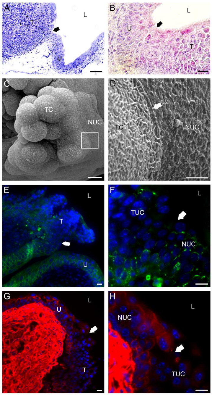
Figure 7.
Characteristics of a bladder tumor margin 3 weeks after intravesical application of MB49-GFP cancer cells. (A) Semi-thin section of a bladder tumor with an easily recognized margin (arrow) between the urothelium (U) and prominent tumor formation (T). Toluidine blue staining. (B) Histological section of bladder tumor with a hardly recognized margin (arrow) between the urothelium (U) and sessile tumor formation (T). H&E staining. (C) Scanning electron micrograph of a papillary tumor with an outlined tumor margin (boxed area). (D) A higher magnification view of the tumor margin (the boxed area from (C)) with well-developed tight junctions (arrow) between a tumor cell (TC) and a normal urothelial cell (NUC). Note the different structures of the plasma membranes of the two cell types. The tumor cell had a typically ruffled plasma membrane, while the urothelial cell had a characteristic scalloped appearance of the apical plasma membrane. (E) The tumor margin (arrow) was revealed due to a positive immunofluorescence reaction against E-cadherin (green fluorescence) among cells of the urothelium (U) and a negative immunofluorescence reaction against E-cadherin among urothelial cells of the tumor formation (T). Note that a weak E-cadherin positivity was present only in deeper urothelial layers of the tumor. (F) A higher magnification view (from E) of the tumor margin (arrow) exposed due to different immunofluorescence patterns of E-cadherin (green fluorescence) in normal NUC than in tumor urothelial cells (TUC). (G) The tumor margin (arrow) was revealed due to positive immunofluorescence reaction against desmoplakin 1/2 (red fluorescence) among cells of the urothelium (U) and negative immunofluorescence reaction against desmoplakin 1/2 among cells of tumor formation (T). Note that desmoplakin 1/2 positivity was present only in deeper urothelial layers of the tumor. (H) A higher magnification view (from (G)) of the tumor margin (arrow) exposed due to different immunofluorescence patterns of desmoplakin 1/2 (red fluorescence) in NUC than in TUC. Nuclei were stained with DAPI (blue fluorescence) in (E–H). TC—tumor cell; L—lumen of the urinary bladder. Bars are 100 µm (A); 20 µm (B); 10 µm (C,E–H); and 2 µm (D).

