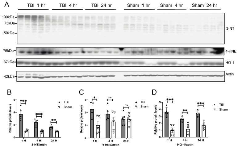
Figure 2.
BOP caused increased oxidative stress in the mouse brain. (A) Representative Western blots demonstrated the early induction of oxidative stress markers in the mouse brain after blast (n = 5/group). Actin was probed as an internal loading control. (B–D) Quantification analysis of protein oxidative marker 3-nitrotyrosine (3-NT) (B), lipid peroxidation marker 4-hydroxynonenal (4-HNE) (C), and antioxidant hemeoxygenase-1 (HO-1) (D) normalized to actin levels. (Data are means ± SEM, * p < 0.05, ** p < 0.01, *** p < 0.001; ns, non-significant).

