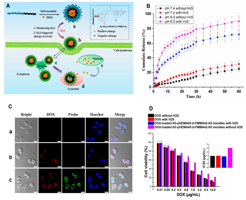
Figure 5.
The mechanism of the drug-loaded hydrogen sulfide-responsive micelles when exposed to the stimulant is depicted schematically (A); the drug release rates of the drug-loaded N3-Nap-pHEMA-b-PMMA-N3 sample at acidic and physiological environments, with/without being exposed to hydrogen sulfide (B); the images of HeLa cells treated with doxorubicin (a), drug-loaded N3-Nap-pHEMA-b-PMMA-N3 without hydrogen sulfide (b), and drug-loaded N3-Nap-pHEMA-b-PMMA-N3 with hydrogen sulfide (c), taken by confocal microscopy (scale bar = 20 μm) (C); the cell viability (%) of drug and non-loaded samples, with/without being exposed to hydrogen sulfide (D). Reprinted from [16], with permission from the American Chemical Society.

