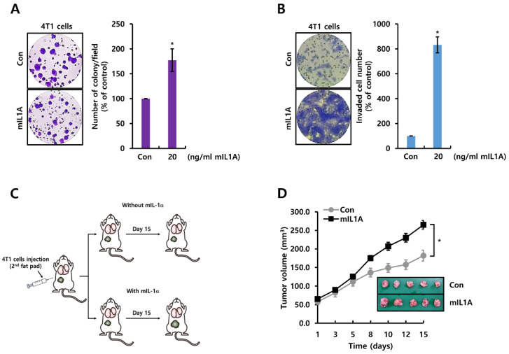
Figure 2.
mIL1A augments cell growth and invasiveness in TNBC cells. (A,B) after cell-seeding, 4T1 TNBC cells were treated with 20 ng/mL mIL1A for two weeks (A) or 24 h (B). Cell proliferation and invasiveness were analyzed by the colony-forming assay (A) and the Boyden chamber assay (B), respectively. The upper compartment of the insert chamber was removed using cotton swabs and the bottom filters were fixed and stained for counting invaded cells. (C) schematic model of the experimental procedure. (D) the size of the tumor in each group (n = 5) was monitored for 15 days. The values are presented as the mean ± standard error of three independent experiments. * p < 0.05 vs. Con. Con; control, mIL1A; mouse IL1A.

