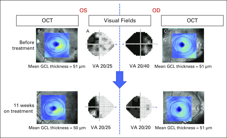
FIG 3.
Improvement in visual fields and acuity, and stable optic nerve structural assessment in subject 2. Ophthalmologic assessment before and 11 weeks after treatment with Debio1347. Humphrey visual fields before therapy (center panels) revealed a combined LHH, from right optic tract involvement, and a bitemporal hemianopia from simultaneous optic chiasmal compression, whereas best-corrected VAs were 20/40 OD and 20/25 OS. Structural assessment of the retinal GCL using OCT demonstrated a thinning of the right retina in both eyes consistent with permanent neuronal loss related to the LHH or optic tract compression (note the lack of normal yellow color on the left side images [B] and [C], reflecting right retinal thinning in the OS and OD, respectively). Mean GCL thicknesses were 51 μm in both eyes. Three months after treatment with Debio1347, repeat visual fields revealed a resolution of the temporal loss OD, reflective of resolved bitemporal hemianopsia, although the LHH persisted, and VAs improved to 20/20 OD and 20/25 OS. OCT demonstrated stable GCL thinning, confirming no further damage to the optic apparatus during that period. Repeat visual fields and OCT 14 months after treatment began (not shown) demonstrated persistent improvement in visual fields and stable structural integrity. GCL, ganglion cell layer; LHH, left homonymous hemianopia; OCT, optical coherence tomography; OD, right eye; OS, left eye; VA, visual acuity.

