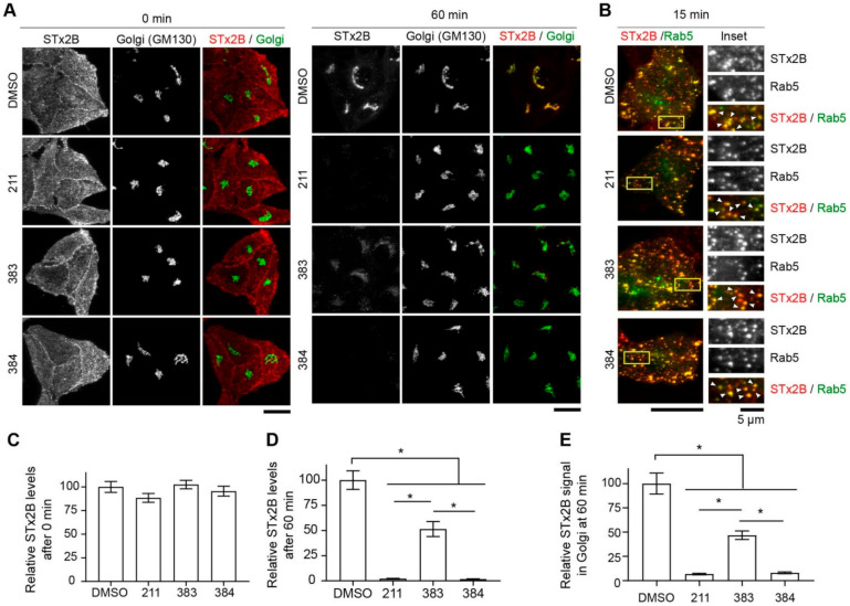
Figure 5.
Compounds 383 and 384 inhibit transport of STx2B to the Golgi. (A). STx2B transport assay in cells treated with DMSO (0.1%) or 10 µM indicated compounds for 24 h. Cells were fixed at 0 or 60 min after the start of transport and processed for microscopy. Scale bars, 25 µm. (B). STx2B transport assay in cells transfected with GFP-Rab5 for 4 h followed by treatment with DMSO (0.1%) or 10 µM indicated compounds for 24 h. Cells were fixed 15 min after the start of transport. Scale bar, 25 µm; inset, 5 µm. Arrows, overlap. (C,D). Quantification of STx2B signal from (A). DMSO-treated cells at 0 min normalized to 100. N ≥ 25 cells per condition. * p < 0.05 by one-way ANOVA and Tukey–Kramer post hoc test for indicated comparisons. (E). Quantification of Golgi-localized STx2B signal from (A). Signal intensity after DMSO treatment at 60 min is normalized to 100. N ≥ 25 cells per condition. * p < 0.05 by one-way ANOVA and Tukey–Kramer post hoc test for indicated comparisons.

