Figure 2.
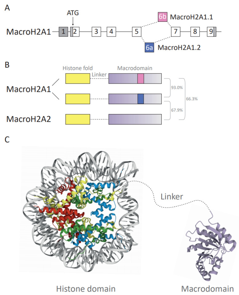
(A) Diagram depicting the structure and splicing of the gene encoding macroH2A1 (H2AFY). Gray boxes represent non-coding exons, white boxes represent coding exons. The macroH2A1.1- and macroH2A1.2-specific exons are in pink and blue, respectively. (B) Schematic of the three human macroH2A variants’ domain architecture. Total amino acid sequence identity is shown as a percentage. (C) Crystallographic structure representation of the macroH2A-containing nucleosome and macrodomains. The protein structure of the unstructured basic linker region depicted in gray is not known. The nucleosome and macrodomain are colored by molecule type. DNA—gray, H2A—yellow, H2B—red, H3—blue, H4—green, macrodomain—purple. The protein structure was generated with protein data bank [70,71] ID: histone fold domain (3REH) [72] and macrodomain of macroH2A1.1 (1YD9) [28].

