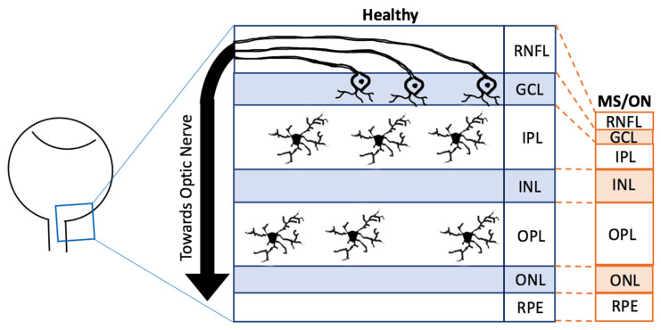
Figure 2.
Comparison of retinal changes in Healthy and Diseased Retinas. A schematic diagram showing changes in the main layers in the healthy (blue) and MS/ON (multiple sclerosis/optic neuritis, orange) retinae. The diagram is not to scale and a hypothetical representation. The ganglion cell axons are found in the RNFL whilst their cell bodies are in the GCL. Microglia are said to reside in the IPL and OPL. There are also other retinal neurons and glia that are not specified in this diagram. RNFL = retinal nerve fibre layer, GCL = ganglion cell layer, IPL = inner plexiform layer, INL = inner nuclear layer, OPL = outer plexiform layer, ONL = outer nuclear layer, RPE = retinal pigment epithelium, MS = multiple sclerosis, ON = optic neuritis.

