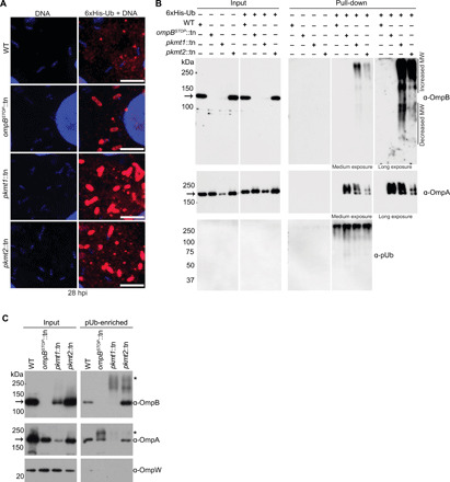
Fig. 3. Lysine methylation protects OMPs from ubiquitylation.

(A) Micrographs of infected Vero cells expressing 6xHis-ubiquitin stained with anti-His antibody (red) and Hoechst (bacterial and host DNA; blue) at 28 hpi (n = 2). Scale bars, 5 μm. (B) Western blot of His-Ub input and pull-down samples from infected control and 6xHis-ubiquitin expressing cells probed for OmpB (affinity-purified anti-OmpB antibody), OmpA (monoclonal anti-OmpA antibody, 13-3), and pUb (FK1, Enzo) (n = 3). (C) pUb-enriched (TUBE-1, pan-specific) samples from purified bacteria probed for OmpB, OmpA as above, and OmpW [anti-OmpW serum; OmpB and OmpA of endogenous molecular weight (MW) represent nonspecific binding to TUBE-1 beads; n = 3]. Asterisks indicate OmpB and OmpA that exhibit increased molecular weight, indicating ubiquitylation. Arrows indicate OmpB and OmpA of endogenous molecular weight.
