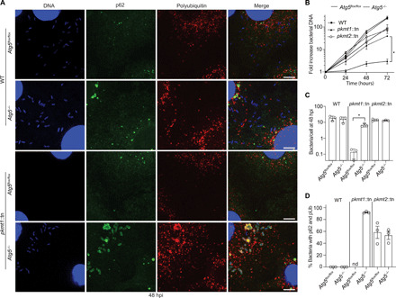
Fig. 7. Methylation prevents ATG5-dependent R. parkeri killing in macrophages.

(A) Micrographs of infected control (Atg5flox/flox) and Atg5-deficient (Atg5−/−) BMDMs at 48 hpi. Fixed with 4% PFA and stained for bacterial and host DNA (Hoechst; blue), polyubiquitin (FK1, Enzo; red), and p62 (anti-p62 antibody; green) (n = 3). Scale bars, 5 μm. (B) Bacterial growth curves in control and Atg5−/− BMDMs, as measured by genomic equivalents using quantitative polymerase chain reaction (PCR; n = 3). (C) Quantification of the mean number of bacteria per cell (n = 3; bacteria from five fields of vision per strain, host genotype, and experiment). (D) Quantification of the percentage of bacteria that colocalize with p62 and pUb (n = 3). nd, not determined. Statistical comparisons for each bacterial strain between the different host genotypes in (B) and (C) were performed using a Brown-Forsyth and Welch ANOVA with Dunnett’s post hoc test. *P < 0.05. All data are means ± SEM.
